
Figure 1 shows what a type A tympanogram looks like. It is located in the 5 oclock position when viewing a normal right tympanic membrane and in the 7 oclock position for a normal left tympanic membrane.
Ear Drum Perforation Ent Clinic

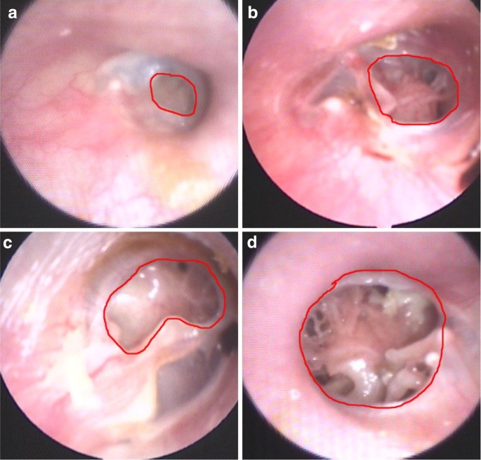
Subtotal Perforation Of Tympanic Membrane Dotted Line Exposing The Download Scientific Diagram

Hole In The Ear Drum Tympanic Membrane Perforation Dr Gan Eng Cern
Consider antibiotic therapy if.
Tympanic membrane perforation. Before covering each below please note that the classification of tympanogram types differs between clinics and audiologists. Pars tensa and pars flaccida The cone of light can be used to orientate. It can also make your middle ear vulnerable to infections.
There are three different types of otitis media associated with a perforation of the tympanic membrane. Perforation of tympanic membrane H72-Code First. A tympanic membrane perforation can have many causes.
The practice of Vernick Gopal welcomes patients with longstanding unusual and unresolved otolaryngology problems. For such conditions ICD-10-CM has a coding convention that requires the underlying condition be sequenced first followed by the manifestation. The tympanic nerve is part of a nerve plexus which transmits impulses that controls salivary secretion.
As an adjunct short-term use of topical analgesia eg 2 lignocaine 1-2 drops applied to an intact tympanic membrane may be effective for severe acute ear pain. X age 6 months x age 2 years with bilateral AOM x symptoms 48 hours x severe symptoms fever 39C and moderate to severe otalgia x evidence of perforation. Complications include otitis media with effusion perforation of the.
Certain conditions have both an underlying etiology and multiple body system manifestations due to the underlying etiology. Click on Pictures to Enlarge. Initial presentation of a young girl.
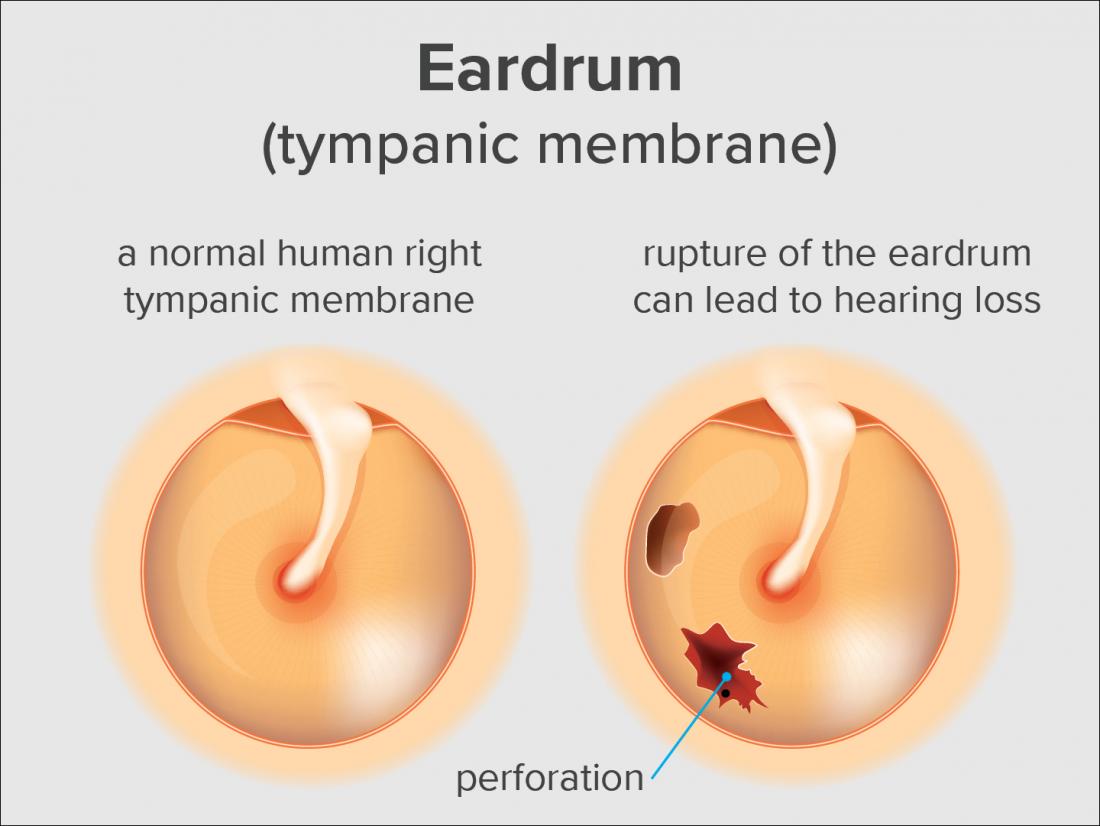
A ruptured eardrum tympanic membrane perforation is a hole or tear in the thin tissue that separates your ear canal from your middle ear eardrum. Lateral process of malleus. X retracted or in the neutral position x amber or blue A fluid level or bubbles may be seen behind the tympanic membrane Box C.
If the perforation is from recent trauma many ear nose and throat specialists will elect to watch and see if it heals on its own. The tympanic membrane is obscured by wax but needs to be viewed to establish a diagnosis. The TM function is to aid in hearing by creating vibrations whenever.
Tympanum tympanic membrane persistent post-traumatic postinflammatory H729- ICD-10-CM Diagnosis Code H729- Unspecified perforation of tympanic membrane. See Diagnosis section for definition. Treatment includes pain control with analgesics and might include antibiotics.
The infection may decompress through a perforation in the tympanic membrane or extend through the lateral mastoid cortex forming a postauricular subperiosteal abscess. These images are a random sampling from a Bing search on the term Tympanic Membrane Perforation Click on the image or right click to open the source website in a new browser window. Search Bing for all related images.
The person wears a hearing aid and an impression needs to be taken for a mould or wax is causing the hearing aid to whistle. Squamous epithelium the skin on the outside of the body has grown into the middle ear through the. Diagnosis is generally made with conventional otoscopy.
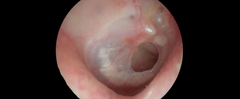
Patterns of Ossicular Deficiency. This picture shows a marginal perforation of the eardrum. Initial management of earwax includes ear drops for 35 days initially to soften wax.
What causes a tympanic membrane perforation. Congenital cholesteatoma rare may appear as a white mass behind an intact tympanic membrane in a person with no prior history of ear discharge tympanic membrane perforation or surgical procedures on the ear. The tympanic membrane or ear drum serves as the window into the middle ear.
The below merely provides an overview of which tympanograms you may come across and what they may indicate. Tympanic membrane perforation also known as a perforated eardrum is a hole in the thin membrane that separates the ear canal from the middle ear. For a normal tympanic membrane you should be able to observe.
Centralpars tensa tympanic membrane perforation with a tympanostomy tube in place. Urgent evaluation by ENT. Rarely it extends centrally causing a temporal lobe abscess or a septic thrombosis of the lateral sinus.
It also serves as the lateral wall of the tympanic cavity separating it from the external auditory canalThe membrane lies across the end of the external canal and. Complicated or Large Perforation. As for simple perforations see above plus.
The term myringoplasty refers to repair of the tympanic membrane alone. In the anatomy of humans and various other tetrapods the eardrum also called the tympanic membrane or myringa is a thin cone-shaped membrane that separates the external ear from the middle earIts function is to transmit sound from the air to the ossicles inside the middle ear and then to the oval window in the fluid-filled cochleaHence it ultimately converts and amplifies vibration in. The TM is a layer of cartilaginous connective tissue with skin on the outer surface and mucosa covering the inner surface that separates the external auditory canal from the middle ear and ossicles.
Direct observation of the tympanic membrane and external auditory canal through an otoscope offers valuable information about possible disease within the middle ear. Isolated Small Perforation Pediatric For perforation due to otitis media PO antibiotics preferred over topical. A ruptured eardrum can result in hearing loss.
Tympanic membrane perforation is when the tympanic membrane TM ruptures creating a hole between the external and middle ear. 1 acute otitis media complicated by perforation of the tympanic membrane. A ruptured eardrum usually heals within a few weeks without treatment.
An eardrum rupture is a small hole or tear in your eardrum or tympanic membrane. The tympanic membrane may be perforated. Outpatient management Complicated or larger perforations require.
Various tympanostomy tube styles and sizes. If there is significant discharge occluding the tympanic membrane referral for examination. There are several options for treating a perforated eardrum.
The tympanic membrane is a thin tissue that divides your middle ear. The membrane may be white yellow pink or red. Tympanic membrane also called eardrum thin layer of tissue in the human ear that receives sound vibrations from the outer air and transmits them to the auditory ossicles which are tiny bones in the tympanic middle-ear cavity.
After 35 combined years in an academic otolaryngology ears nose and throat practice David Vernick MD and Harsha Gopal MD decided to form a private practice in 2004 with a commitment to provide expert and timely care to patients in the greater Boston area. Decongestants antihistamines and corticosteroids are not effective in AOM.
Ruptured Eardrum Symptoms Causes And Treatments

Tympanic Membrane Rupture Wikem
1

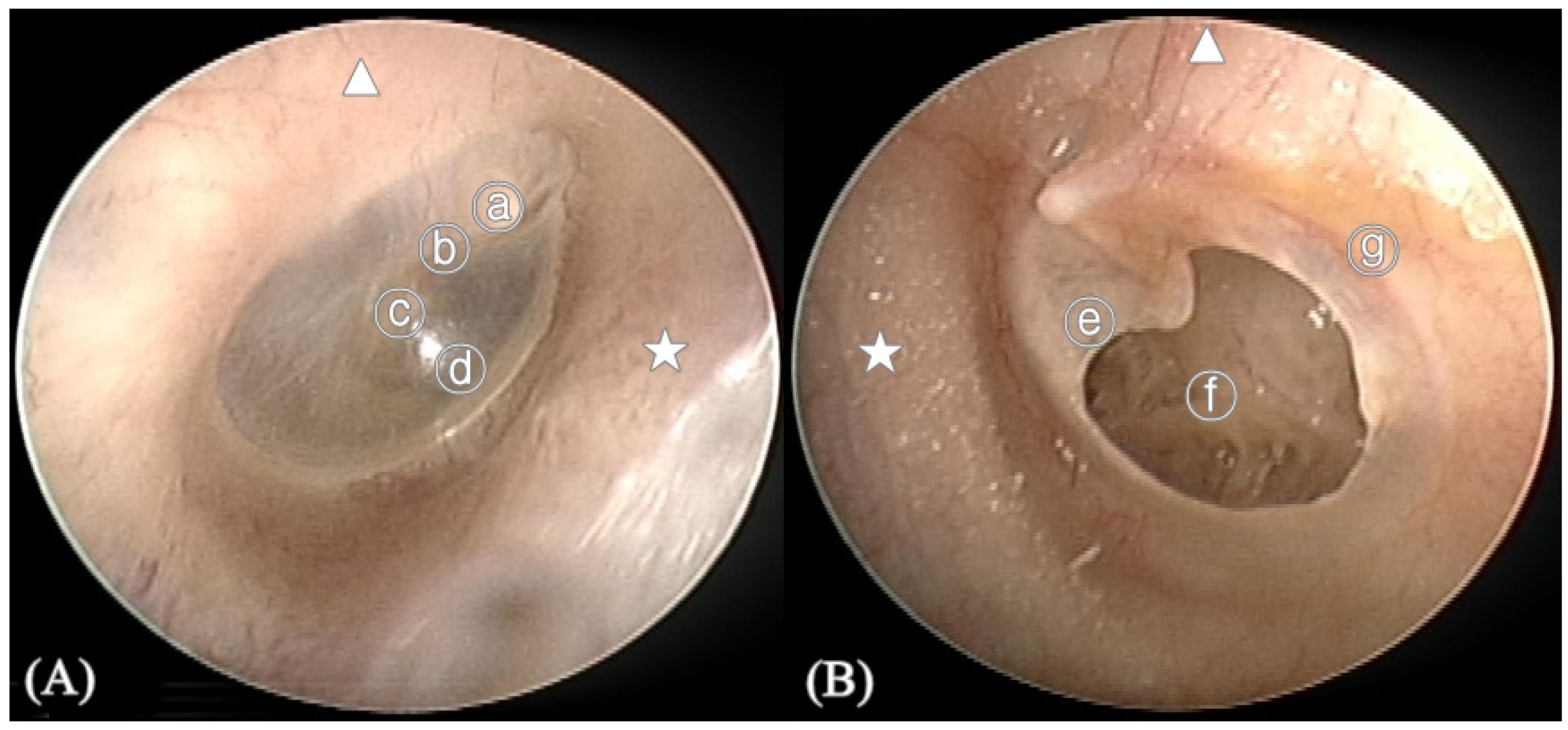
Normal And Perforated Tympanic Membrane A Normal Right Tympanic Download Scientific Diagram
Applied Sciences Free Full Text Automated Classification Of The Tympanic Membrane Using A Convolutional Neural Network Html
Assessment Of Hearing Loss Induced By Tympanic Membrane Perforations Under Blast Environment Springerlink

Eardrum Rupture Otolaryngology Specialists Of North Texas
Tympanic Membrane Perforation A Hole In The Ear Drum And Tympanoplasty Myringoplasty Dr Sean Flanagan


